Comprehensive Guide to Double Chin Surgery: Why Anatomical Diagnosis and Deep Neck Lifts Matter
If you are struggling with a persistent double chin, submental fullness, or neck laxity, you have likely come across various anti-aging solutions, with double chin surgery frequently recommended. However, achieving a truly sharp, defined jawline requires identifying the precise anatomical cause of your submental fullness and choosing the right surgical approach accordingly.

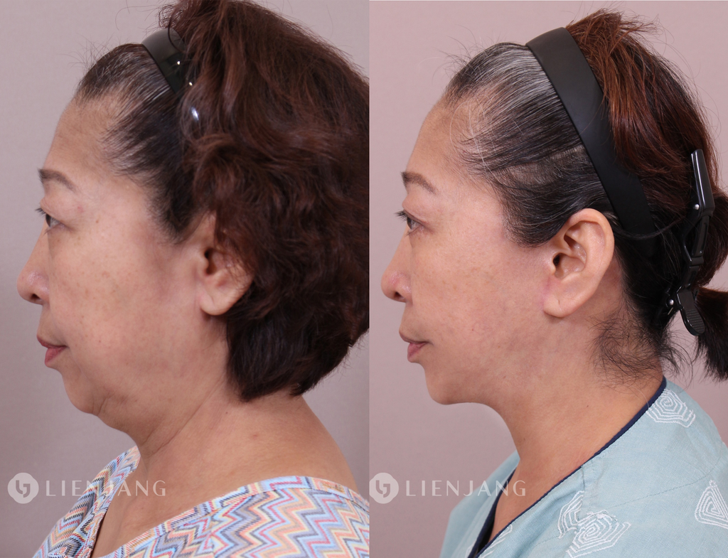
The Core Anatomy: What Causes a Double Chin?
Anatomically, a double chin is driven by multiple overlapping layers:
- Skin elasticity and laxity
- Subcutaneous fat distribution
- Platysma muscle integrity
- Deep fat compartments beneath the platysma
- Digastric muscle volume
- Submandibular gland size and positioning
- Secondary skeletal and postural elements (such as jawbone shape, hyoid bone height, and forward head posture)
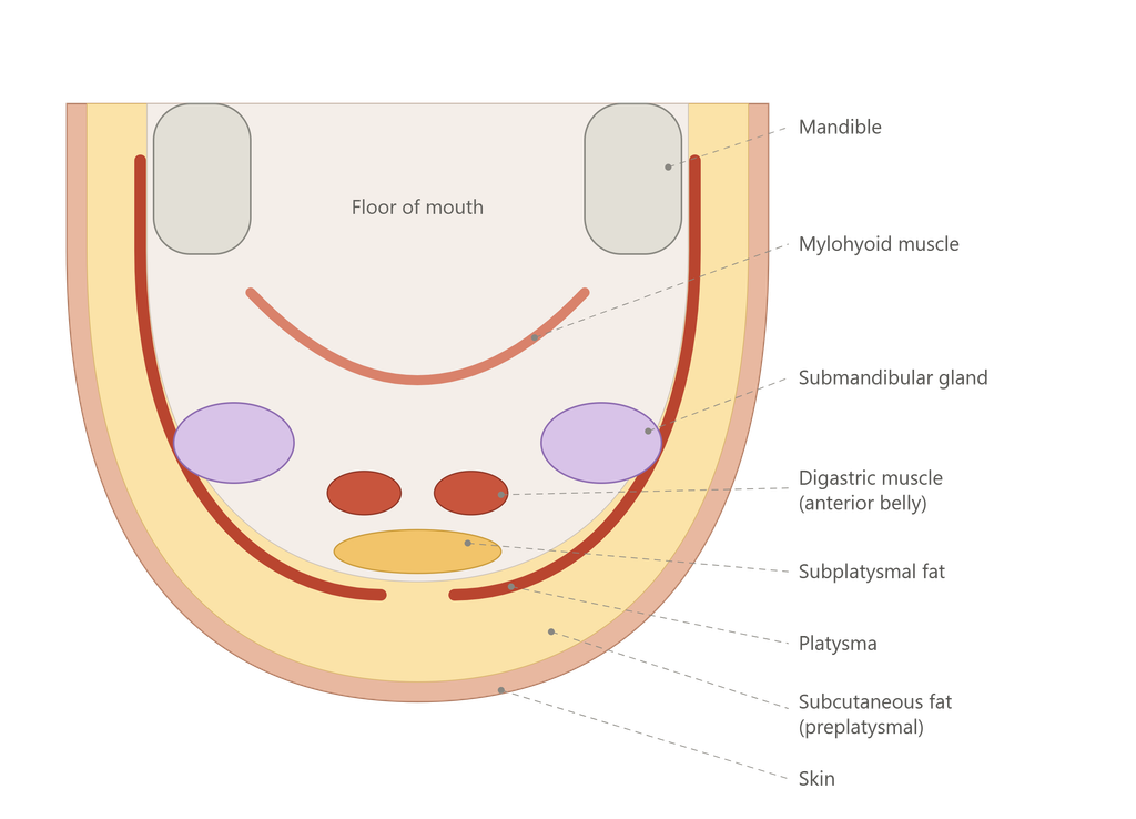
Because no two necks age or develop the exact same way, an accurate structural evaluation is critical. This is why ultrasound diagnostic imaging is indispensable in modern facial rejuvenation.
The Role of Ultrasound Diagnosis in Customizing Your Surgery
Relying on a physical exam alone is often insufficient for mapping out submental anatomy. By utilizing advanced ultrasound diagnostics, we can evaluate what is truly happening beneath the surface:
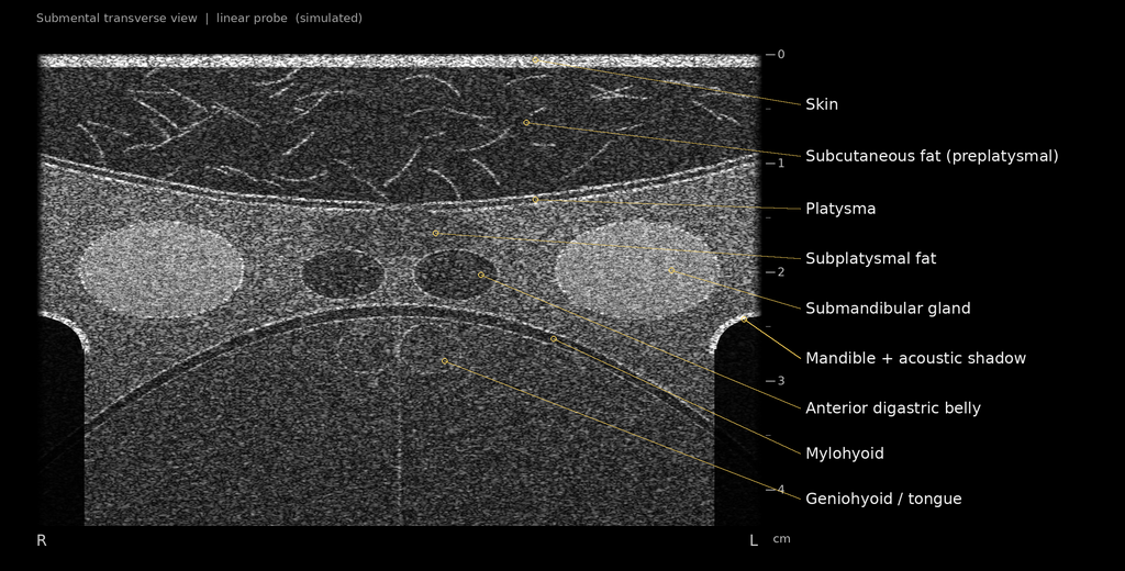
Skin: The bright (hyperechoic) topmost band.
Subcutaneous Fat: Dark (hypoechoic) background scattered with bright fibrous septae (thickened in fat-dominant double chins).
Platysma Muscle: Bounded by two bright parallel fascial lines with a central midline gap.
Subplatysmal Fat: Located directly beneath the platysma with typically blurred margins.
Digastric Muscle (Anterior Belly): Two dark oval structures situated on either side of the midline.
Submandibular Gland: Uniformly bright oval masses on both sides, appearing noticeably more echogenic than muscle.
Mylohyoid Muscle: The deeper arch-shaped dark band.
Mandible: Bright bony contours at the outer edges casting posterior acoustic shadowing.
- Fat Layer Thickness: Distinguishing between superficial subcutaneous fat and deep subplatysmal fat.
- Muscle Laxity: Assessing whether the platysma muscle is separated, weakened, or sagging.
- Glandular and Muscular Enlargement: Determining whether the submandibular glands or digastric muscles are prominent and contributing to fullness.
Once we identify the exact underlying drivers through ultrasound, we can tailor the surgical strategy to your unique anatomical profile.
Tailored Surgical Strategies by Anatomical Cause
1. Skin Laxity
- Younger Patients: Creating deep, defined subangular contours allows the skin to redrape naturally, minimizing the appearance of laxity without major excisions.
- Mature Patients with Excess Skin: For patients with significant skin redundancy, excising excess skin combined with an lateral/extended neck lift is necessary to restore smooth cervical contours.

2. Excess Subcutaneous Fat
- When the primary issue is localized fat just beneath the skin, targeted liposuction, energy-based devices, or lipolytic treatments can successfully reduce volume.
3. Platysma Muscle Separation (Cervical Webbing)
- If the platysma muscles have separated or sagged, they must be sutured and secured midline—often referred to as a corset platysmaplasty—to rebuild a strong, taut structural sling under the jaw.
4. Deep Structural Volume (Subplatysmal Issues)
- The Crucial Distinction: Superficial liposuction and non-invasive energy devices cannot reach or reduce deep-layer volume. If your double chin is caused by deep anatomical structures, surgical reduction is mandatory.
- Identifying Deep Volume:
- Digastric Muscle: If pressing your tongue against the roof of your mouth while moving your submental musculature causes a central bulge, the digastric muscle is often a primary contributor.
- Submandibular Glands: If firm, prominent masses are palpable and visible along the lateral jawline, enlarged submandibular glands are likely the cause.
- Deep Fat Pockets: Discrete fat deposits situated underneath the platysma.
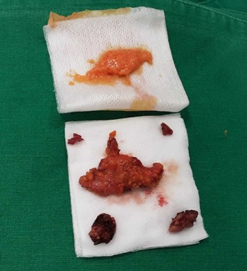
- Deep Neck Dissection: To resolve these deep-seated issues, a specialized deep neck dissection is required. This involves precise excision of deep fat pockets, partial reduction or contouring of the digastric muscles, and partial sculpting of the submandibular glands.
Frequently Asked Questions (FAQ)
Q1. Why didn’t my previous double chin liposuction or thread lift work?
A: Liposuction and non-invasive tightening devices only treat superficial subcutaneous fat and skin laxity. If your double chin is caused by a separated platysma muscle, bulky digastric muscles, or enlarged submandibular glands, superficial treatments will yield little to no visible improvement. A comprehensive anatomical evaluation using ultrasound is necessary to identify and treat these deep-layer components.
Q2. What is a Deep Neck Lift (or Deep Neck Dissection)?
A: A deep neck lift goes beyond standard skin tightening and superficial liposuction. It addresses the anatomical structures located beneath the platysma muscle. This may include excising deep fat compartments, managing enlarged submandibular glands, and contouring the digastric muscles to create a deeply sculpted, sharp cervicomental angle.
Q3. How do I know if my double chin is caused by muscle or fat?
A: During a clinical consultation, high-resolution ultrasound imaging is used to visualize your internal neck anatomy in real-time. Additionally, physical maneuvers—such as flexing the tongue against the upper teeth—help differentiate whether a submittal protrusion is driven by skin, fat, the platysma, the digastric muscle, or the salivary glands.
Q4. Can submandibular glands safely be reduced during neck surgery?
A: Yes, when performed by an experienced plastic surgeon specializing in facial rejuvenation, partial reduction of the submandibular glands is a safe and highly effective technique for eliminating lateral fullness along the jawline while preserving normal saliva production.
Q5. Who is an ideal candidate for double chin surgery or a neck lift?
A: Ideal candidates are individuals bothered by persistent submental fullness, undefined jawlines, neck bands, or skin laxity that does not respond to weight loss or non-invasive treatments. A personalized surgical plan is essential to match the procedure to your specific anatomical findings.



Medical Disclaimer:The content provided on this blog is for informational and educational purposes only and does not constitute medical advice, diagnosis, or treatment. Individual surgical results may vary, and all medical or surgical procedures carry inherent risks. Always seek the advice of a qualified healthcare provider with any questions regarding a medical condition or treatment options.