[Case Review] Total Anti-Aging in a Patient in Her 60s: From Endoscopic Forehead Lift to Deep Plane Facelift and Deep Neck Lift
[Notice on Clinical Documentation and Copyright Protection] All clinical photographs and pre/post-operative recovery records utilized in this post are published under the voluntary and explicit informed consent of the patient, in strict compliance with medical and privacy laws. Unauthorized duplication, copying, distribution, or commercial reuse of any images and text content contained within this blog/website is strictly prohibited and subject to legal liability.
📋 Executive Summary
This comprehensive anti-aging complex surgery case review details the successful correction of multi-planar facial aging in a female patient in her 60s (featuring forehead wrinkles, upper eyelid hollowness, deep nasolabial folds, recurrent lower eyelid fat herniation, blunted jawline, and cervical laxity). Rather than relying on isolated interventions, this case utilized an anatomically targeted, multi-modality surgical strategy:
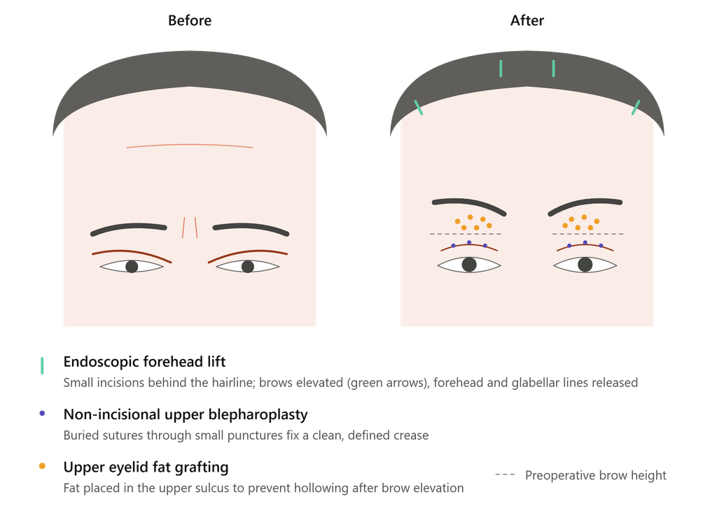
- Upper Face: Endoscopic Forehead Lift + Non-Incisional Upper Blepharoplasty + Upper Eyelid Fat Grafting (correction of forehead wrinkles and periorbital drooping, eyelid fold reinforcement, and hollowness prevention)
- Mid-to-Lower Face: Deep Plane Facelift (structural release of retaining ligaments to soften deep nasolabial folds and tear troughs / Indian bands)
- Lower Face & Neck: Deep Neck Lift with Platysmal Hammock Technique (sculpting of an irregular jawline, correction of neck sagging, and smoothing of horizontal neck rings)
- Lower Eyelids: Lower Blepharoplasty Revision (refining a prior surgical scar and redistributing herniated orbital fat)
- Progress & Findings: Marked structural elevation and stable scar/ear morphology (free of distortion) were confirmed by the 1-month post-op mark following an acute recovery phase at week 1.
- Note on limitations: Due to variable graft take, a minor recurrence of upper eyelid hollowness was observed. A secondary touch-up fat injection is planned to ensure a realistic and transparent clinical outcome.
1. Pre-operative Patient Analysis
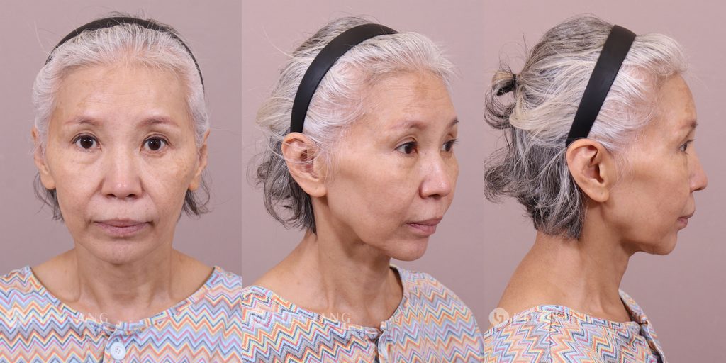
A female patient in her 60s presented with extensive, structural facial aging accompanied by complex functional and aesthetic complaints:
- Upper Face (Forehead & Periorbital Region): Severe upper eyelid dermatochalasis forced the patient into an unconscious compensatory frontalis over-pull, resulting in deeply fixed, horizontal forehead wrinkles and a compressed, narrow forehead presentation. Weak pre-existing double eyelid folds and an underlying involutional upper eyelid hollowness created a structural hazard where an isolated forehead lift would aggravate the hollow look and disrupt crease integrity.
- Mid-to-Lower Face (Nasolabial Folds & Philtrum): Deep nasolabial folds combined with soft tissue and retaining ligament descent, making her lateral philtrum appear elongated and prematurely aged. Overall midface volume depletion and prominent Indian bands were noted.
- Lower Face & Neck (Lower Eyelids, Jawline & Neck): A history of prior lower blepharoplasty left a noticeable scar alongside re-herniated orbital fat. The mandibular border was blunted and irregular, accompanied by distinct cervical skin laxity and deep horizontal neck rings.


2. Surgical Plan and Strategic Approach
By analyzing individual aging vectors and precise facial fascial planes, a harmonized multi-vector surgical plan was executed:
- Endoscopic Forehead Lift + Non-Incisional Upper Blepharoplasty + Upper Eyelid Fat Grafting: Released and smoothed forehead and glabellar creases while repositioning descended eyebrows to an optimal youthful height. A non-incisional blepharoplasty secured clean crease definition, and concurrent upper eyelid fat grafting was performed to help prevent postoperative upper eyelid hollowing.
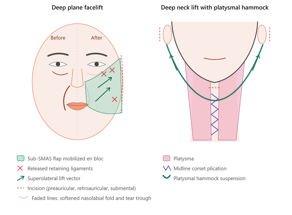
- Deep Plane Facelift: Accessed beneath the superficial musculoaponeurotic system (SMAS) layer to thoroughly release deep retaining ligaments (including zygomatic ligaments). This mobilized the facial soft tissues en bloc to soften tear troughs and reposition deep nasolabial folds superiorly and laterally, restoring a shortened, vibrant philtrum length.
- Deep Neck Lift with Platysmal Hammock Technique: Addressed submental fullness and cervical laxity via micro-incisions behind the ears and submental approach. The platysma muscle was corseted and cradled using a platysmal hammock technique, refining the jawline into a crisp V-line and lifting deep cervical contours.
- Lower Blepharoplasty Revision: Excised the conspicuous prior scar and meticulously redistributed/secured re-herniated orbital fat to re-establish a smooth, natural lower eyelid contour.
3. Post-operative Progress and Timeline Analysis

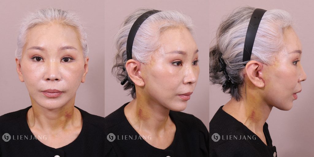

- 1 Week Post-operative (Acute Recovery Phase): Because a comprehensive, multi-planar intervention spanning the forehead, periorbital structures, face, and neck was performed, bruising and edema (particularly along 45-degree lateral vectors and submental lymphatic pathways) peaked during this period. While tissue healing introduced tightness, foundational corrections—such as the lifting of eyelid heaviness, flattening of nasolabial folds, and elevation of sagging jowls—were already securely in place.
- 1 Month Post-operative (Early Result & Scar Stabilization Phase): As residual edema subsided, the structural benefits of the deep plane and endoscopic forehead lifts became prominent. In the 45-degree view, nasolabial depths and Indian bands were markedly reduced, and the jawline and neck angle achieved clean definition.
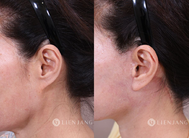
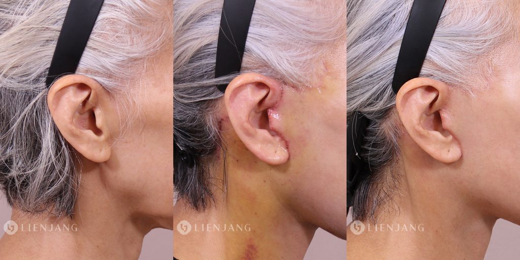
- Ear Morphology & Scarring: Preauricular and postauricular incision lines showed minimal visibility and stable healing by week 4 due to precise, tension-free closure. Crucially, there was no structural ear distortion (such as a pixie ear), preserving natural sideburn and ear contour anatomy.
- Graft Take Limitation: While the core upper facial framework healed impeccably, the initial upper eyelid fat graft exhibited a somewhat lower graft take rate, leading to a slight recurrence of upper eyelid hollowness at the 1-month mark. A simple, secondary micro-fat touch-up is planned to achieve ideal long-term fullness.
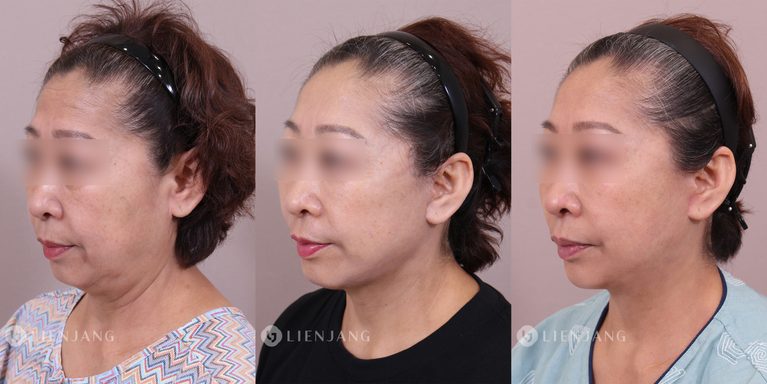
4. Angle-Specific Clinical Efficacy Analysis: Pre-op vs. 1-Week vs. 1-Month Progression
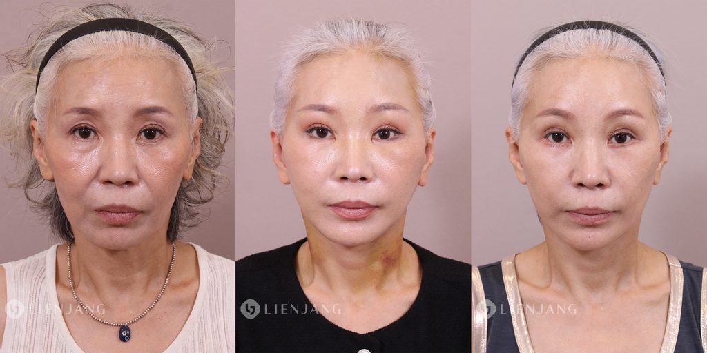
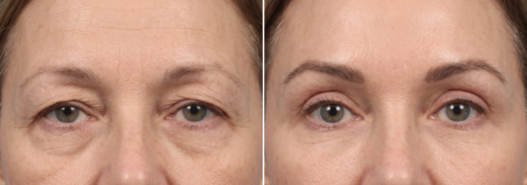
Center (1 Week Post-op): Acute recovery phase with expected swelling; immediate vertical elevation of brows and opening of the upper periorbital area.
Right (1 Month Post-op): Subsided edema revealing a refreshed, symmetric facial frame with smoothed forehead creases and stable double eyelid folds.
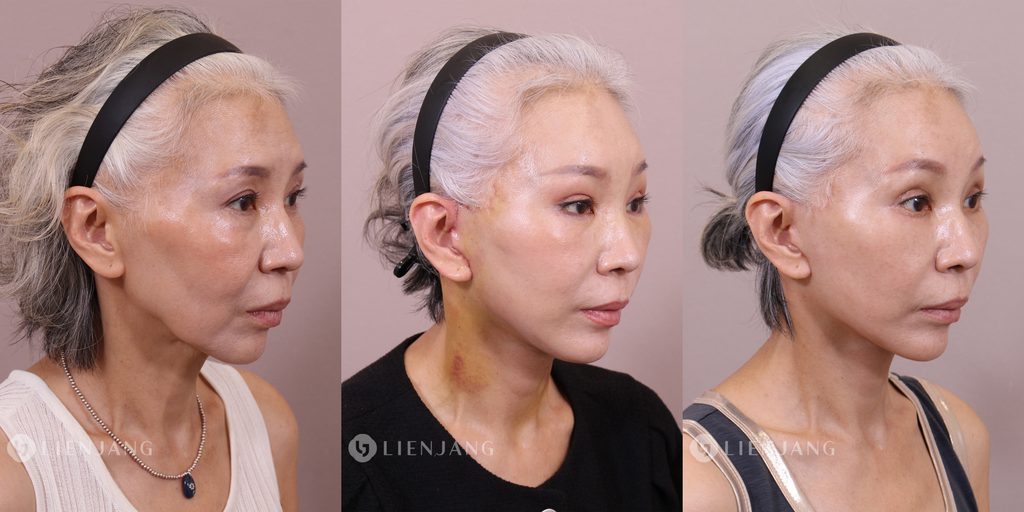
Left (Pre-op): Pronounced Indian bands (tear troughs), deep nasolabial folds, and clear indicators of midface descent.
Center (1 Week Post-op): Peak zygomatic and cheek edema, with the deep plane lift vector securely maintaining upward tissue suspension.
Right (1 Month Post-op): Remarkable transformation showing deep nasolabial folds and Indian bands successfully softened into natural, youthful cheek contours.
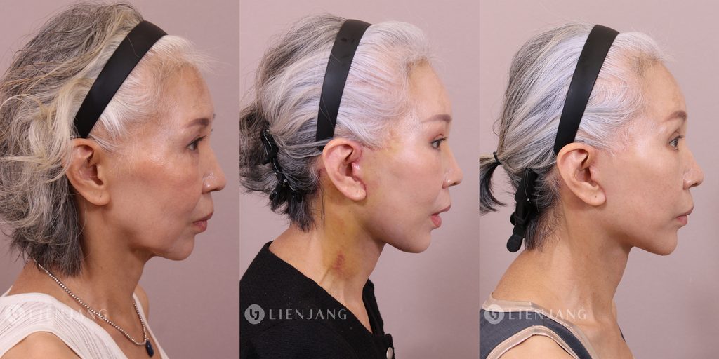
Left (Pre-op): Blunted, irregular mandibular border, submental laxity, and prominent horizontal cervical rings (neck sagging).
Center (1 Week Post-op): Initial structural tension and submental contouring observed beneath post-op swelling.
Right (1 Month Post-op): Crisp, well-defined V-line jaw contour and an elegant, sharp neck angle achieved via the Deep Neck Lift and platysmal hammock technique.
Evaluating the surgical outcomes from multiple angles highlights how the structural corrections progressively manifest from the acute recovery phase to early stabilization
- Frontal View
- Pre-op: Dominated by deep horizontal forehead lines driven by frontalis muscle compensation, a compressed upper face, heavy dermatochalasis, prominent tear troughs, and an aged, elongated lower face.
- 1 Week: Residual diffuse swelling and ecchymosis obscure fine definitions, but the dramatic vertical lifting effect is immediately apparent—eyebrows are elevated, the upper eyelids feel open, and jowl heaviness is significantly reduced.
- 1 Month: Swelling subsides to reveal a symmetric, refreshed frontal frame. The forehead lines are smoothed via endoscopic release, the double eyelid fold is clean and stable, and the midface regains balanced proportions with a shortened, youthful philtrum appearance.
- 45-Degree View
- Pre-op: The 45-degree angle typically highlights the deep depression of Indian bands (tear troughs / cheek-lid junctions), prominent nasolabial folds, and the transition point where facial sagging begins.
- 1 Week: Peak edema is evident along the zygomatic and submandibular vectors. However, the vector shift from the deep plane lift is already holding the tissues securely upward and laterally.
- 1 Month: This angle demonstrates the true triumph of the Deep Plane Facelift. The sharp demarcation of the Indian bands is thoroughly softened, the deep nasolabial folds are smoothly transitioned into natural cheek contours, and the mid-cheek fullness is successfully restored.
- Lateral Profile View
- Pre-op: Characterized by a blunted mandibular border, submental laxity, and pronounced horizontal neck rings / cervical sagging.
- 1 Week: Tightness and submental contouring are visible beneath protective dressings and initial swelling, confirming that the structural framework has been repositioned.
- 1 Month: The Deep Neck Lift with the platysmal hammock technique reveals its defining aesthetic benefit. The jawline is sharply demarcated into a crisp V-line contour, the submental angle is cleanly defined without skin redundancy, and the neck lines are drastically smoothed out, delivering an elegant profile angle.
FAQ Section
Q. Is the recovery period too long when combining an endoscopic forehead lift and a deep plane facelift in patients in their 60s?
A. Combining an endoscopic forehead lift with a deep plane facelift and neck lift naturally extends the surgical scope, leading to concentrated bruising and swelling during the first week. However, because the tissue vectors are precisely managed and structural release is performed along natural fascial planes, major swelling typically begins to subside significantly by week 1. Patients can generally resume social activities and natural facial expressions around the 1-month mark.
Q. Can a facelift cause pixie ear deformity or unnatural tension around the earlobes?
A. Pixie ear or earlobe distortion typically occurs when excessive tension is placed directly on the skin during closure. A deep plane facelift relocates facial soft tissues at the deep muscular layer (SMAS) and retaining ligament level rather than pulling on the skin. This enables a tension-free closure that fully preserves the natural anatomy of the sideburns and ears.
Q. Why choose an endoscopic forehead lift instead of traditional upper blepharoplasty or a direct brow lift?
A. Traditional upper blepharoplasty or direct brow lifts involve direct incisions below the brow or along the eyelid crease, which can leave visible scars and sometimes yield an unnatural, surprised expression. An endoscopic forehead lift utilizes tiny hidden incisions behind the hairline to vertically elevate the entire brow and forehead complex, effectively resolving upper lid hooding and releasing fixed frown lines while keeping scars completely hidden.
Q. Where are the scars located after a Deep Neck Lift with the platysmal hammock technique?
A. The deep neck lift and platysmal hammock procedure are performed through discreet micro-incisions hidden beneath the chin and behind the ears (retroauricular area). While mild pinkness may be visible initially, these incision lines fade naturally over time, blending smoothly into the natural skin creases and remaining virtually undetectable.
Q. How do you prevent upper eyelid hollowing when performing a forehead lift in older patients?
A. Elevating the eyebrows can sometimes accentuate pre-existing upper eyelid hollowing or sulcus depression due to tissue displacement. To prevent this, our surgical strategy incorporates concurrent upper eyelid micro-fat grafting during the endoscopic lift procedure, ensuring balanced volume replenishment and maintaining a smooth, youthful periorbital contour.


Medical Disclaimer:The content provided on this blog is for informational and educational purposes only and does not constitute medical advice, diagnosis, or treatment. Individual surgical results may vary, and all medical or surgical procedures carry inherent risks. Always seek the advice of a qualified healthcare provider with any questions regarding a medical condition or treatment options.