Comprehensive Guide to Endoscopic Forehead Lift: Incisions, Dissection, Fixation, and Before & Afters
1. Introduction: Beyond Simple Eyebrow Elevation
“My eyes feel heavy and fatigued all the time.”
“My eyebrows have dropped, making me look stern or upset.”
“I’m unsure if getting Botox every few months is the right long-term solution…”
These are among the most frequent concerns patients bring to my consultation room.
When your eyes look tired or heavy, the underlying issue is often not just sagging eyelid skin, but a fundamental structural change in the forehead and brow position.
Endoscopic Forehead Lift (or Endoscopic Brow Lift) was specifically developed to address this structural shift. Performed through minimal incisions, this advanced procedure does much more than simply “pull up” the eyebrows—it precisely resets and harmonizes the entire upper facial architecture.
In this article, we will examine the anatomical foundation, surgical techniques, fixation options, and recovery process of an endoscopic forehead lift.
2. Anatomical Background: The Forehead and Eyebrows Function as a Single Unit
To achieve natural, long-lasting rejuvenation, a surgeon must treat the forehead and brows as an integrated structural system.
2.1 The 5 Layers of the Forehead
Understanding the tissue layers is key to understanding surgical safety and effectiveness:
- Skin
- Cubcutaneous tissue (Fat layer)
- Aponeurosis / Galea aponeurotica (A thin, dense fascial sheet connecting frontalis and occipitalis muscles)
- Loose areolar tissue (The primary avascular plane for surgical dissection)
- Periosteum (The dense connective tissue covering the frontal bone)
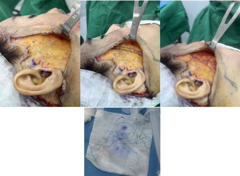
During an endoscopic brow lift, dissection typically takes place in either the subgaleal or subperiosteal plane. Operating along these deep planes provides a safe environment while allowing extensive mobilization of sagging tissues.
2.2 Muscle Mechanics: The Brow Elevators vs. Depressors
The position of your brows is governed by a delicate balance between competing muscle groups:
- Brow Elevator: Frontalis muscle (lifts eyebrows upward)
- Brow Depressors: Corrugator supercilii, Procerus, and Orbicularis oculi (pull eyebrows downward and cause frown lines)
As we age, the downward pull of the depressor muscles naturally overcomes the weakening elevator muscle. This imbalance leads to brow ptosis, hooded eyelids, and deep horizontal or vertical forehead furrows.
3. Endoscopic Forehead Lift Technique: Minimal Incisions, Maximum Precision
3.1 Incision Sites
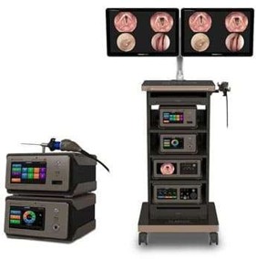
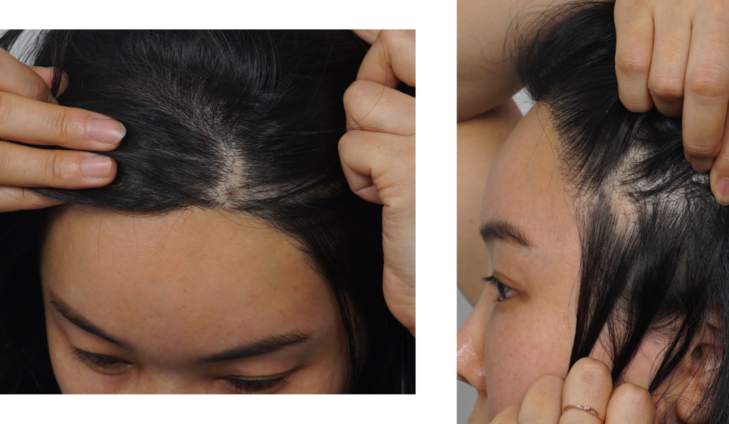
Using endoscopic cameras and specialized instruments, the surgery requires only 4 small incisions (approx. 1 cm each) hidden well within the hairline:
- Two Paramedian Incisions: Positioned on the upper forehead to release and lift the central brow.
- Two Temporal Incisions: Positioned in the temple region to lift the lateral tail of the eyebrow.
Because these incisions are concealed entirely within the hair-bearing scalp, visible scarring is minimal, and healing is rapid.
3.2 Extent of Dissection
- Central Forehead: Dissection proceeds down to the glabella and orbital rim to adjust the inner brow position.
- Lateral Forehead: Follows the temporal fascia to release lateral brow hooding around the outer eye.
- Arcus Marginalis & Supraorbital Rim: Complete release of restrictive ligamentous attachments is essential for true, tension-free elevation.
3.3 Muscle Modification
- Corrugator & Procerus Muscles: Partially and selectively resected or weakened to eliminate dynamic frown/glabellar lines.
- Lateral Orbicularis Oculi: Gently relaxed to relieve crowding around the outer eyes.
- Frontalis Muscle: Preserved intact to maintain natural forehead mobility and expression.
4. Fixation Methods: Securing Long-Term Results
Elevating tissue is only half the battle; ensuring stable, long-lasting fixation is what defines a successful surgery. Several clinical techniques are available, each tailored to individual patient anatomy:
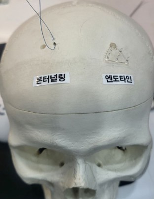
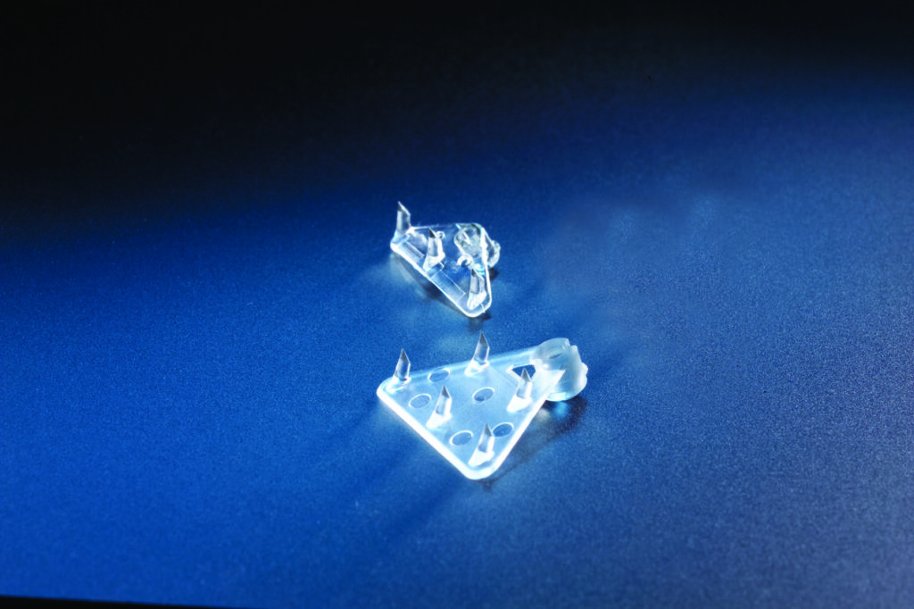
| Fixation Method | Description | Primary Advantages | Key Considerations |
| Endotine / Fixtine | Bioabsorbable multi-point fixation device anchored into cortical bone. | Easy force distribution; highly reliable symmetrical elevation. | Patients with very thin skin may temporarily feel the device underneath. |
| Bone Tunneling | Creating tiny tunnels in the outer bone cortex to secure tissues via sutures. | Zero foreign body feel; highly natural integration. | Technically demanding; requires micro-drilling. |
| Fixtine Screw | Small bioabsorbable screw anchor. | Low-profile fixator; minimal palpability. | Fixation strength relies heavily on cortical bone quality. |
| Gliding Lift / Net | Dermal and subcutaneous vector suture fixation without bone drilling. | Avoids bone alteration entirely; minimal invasiveness. | Lower structural longevity; primarily suitable for mild laxity. |
Clinical Insight: There is no single “best” fixation technique. The choice of device or method depends entirely on the patient’s forehead height, scalp thickness, bone density, hair density, and aesthetic goals.
5. Recovery Timeline & Potential Risks
Expected Recovery Timeline
- Suture / Staple Removal: 7 to 10 days post-op
- Resolution of Bruising & Swelling: ~2 weeks
- Return to Work / Light Activity: 3 to 5 days
- Strenuous Exercise / Sauna: 2 to 3 weeks
Potential Side Effects
- Scalp Numbness / Itchiness: Temporary sensory alterations around the scalp are common and usually resolve within a few weeks to months as sensory nerves recover.
- Postoperative Nausea / Dizziness: Mild, transient nausea may occur in the first 24–48 hours due to swelling or endoscopic manipulation.
- Asymmetry: Asymmetric postoperative swelling can temporarily mimic uneven brow height, which normalizes as tissue heals.
6. Conclusion: Re-Architecting Facial Harmony
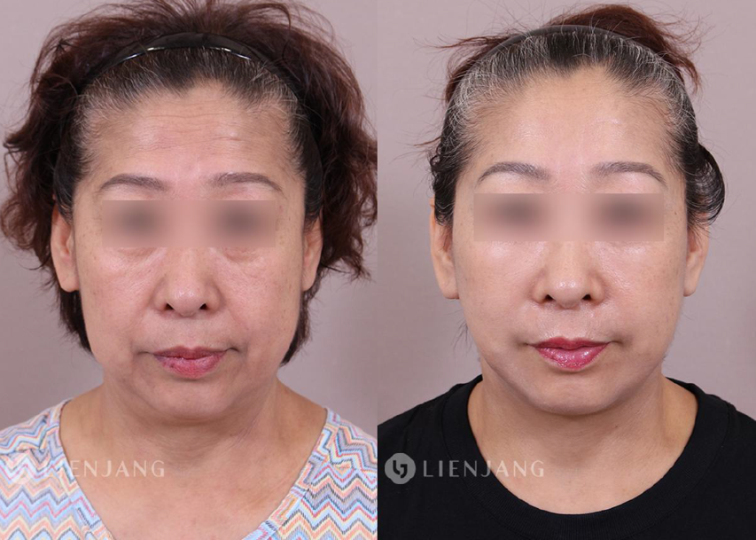
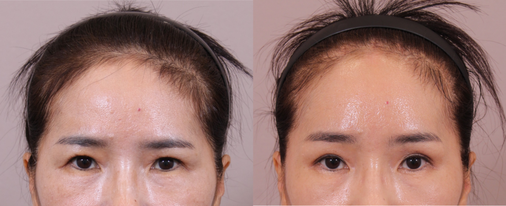
An Endoscopic Brow Lift is far more than a cosmetic fix for sagging eyebrows—it is an anatomical reset of the upper third of the face.
- It addresses heavy, hooded eyes without altering your natural eyelid crease unnecessarily.
- It softens angry or tired expressions caused by hyperactive depressor muscles.
- It achieves meaningful rejuvenation through hidden, minimal incisions with rapid recovery.
If you feel your gaze has grown heavy or tired over time, evaluating the structural relationship between your forehead and brows is the most definitive step toward restoring a refreshed, natural expression.

Medical Disclaimer:The content provided on this blog is for informational and educational purposes only and does not constitute medical advice, diagnosis, or treatment. Individual surgical results may vary, and all medical or surgical procedures carry inherent risks. Always seek the advice of a qualified healthcare provider with any questions regarding a medical condition or treatment options.